10000 Instagram Followers | Boost Your Page Credibility | Fast, Safe & No Password Needed
Ready to grow your Instagram and stand out from the crowd? 📈
This service is perfect for page owners, small businesses, content creators, and entrepreneurs who want real Instagram growth quickly, safely, and without any complicated steps.
📦 What’s Included:
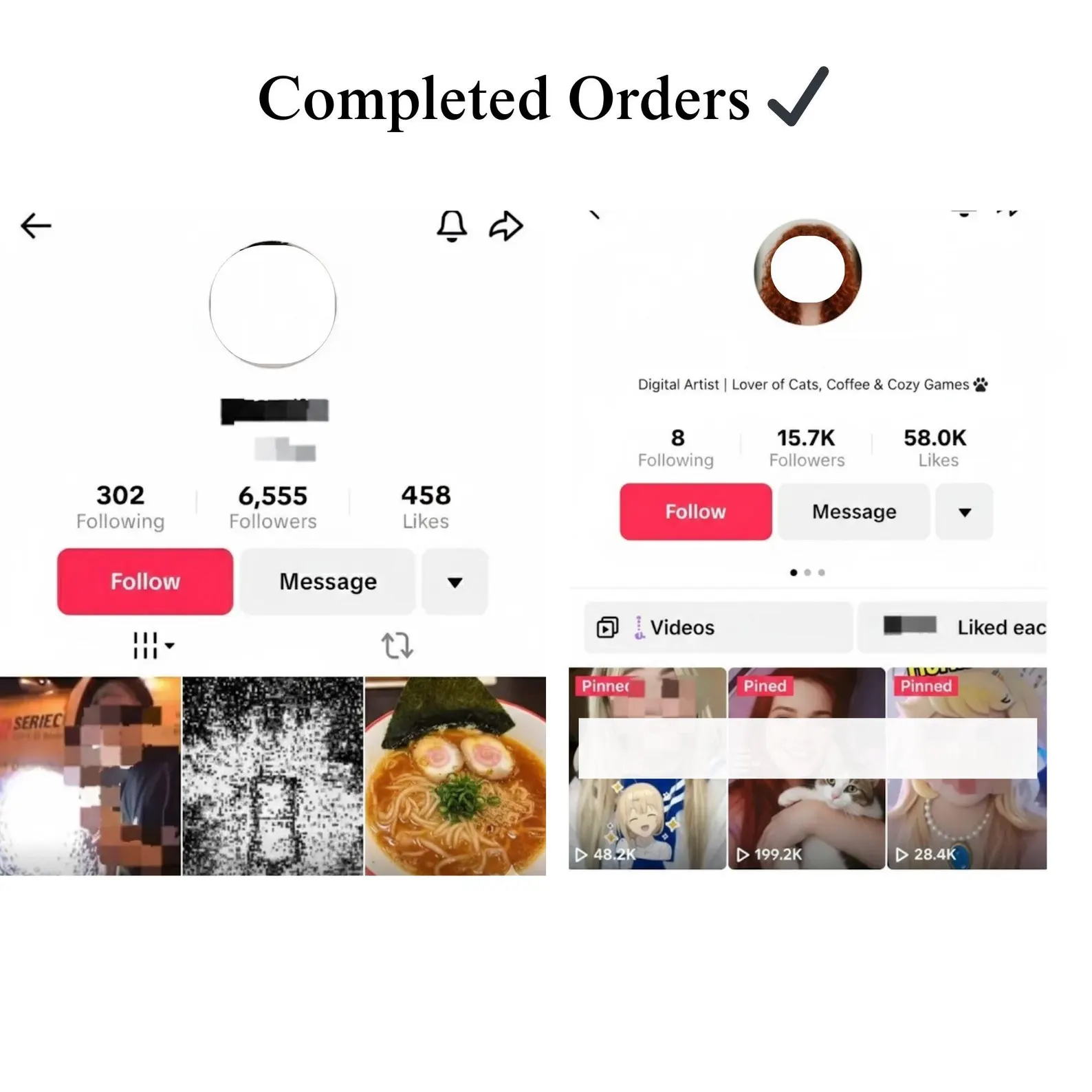
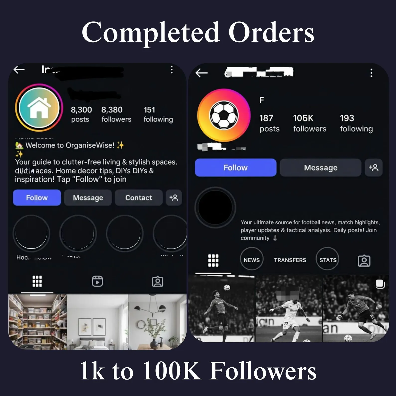
✔️ 10000 Instagram Page Followers
✔️ Gradual, natural-looking delivery
✔️ No password or login required ever
✔️ 100% digital service fast & hassle-free
⚙️ How It Works:
🛒 Purchase this listing
✅ Make sure your account is set to Public
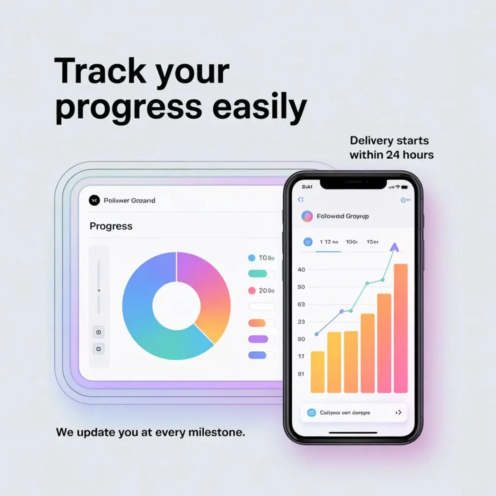
⏳ Delivery begins within 12-24 hours
📈 Watch your followers and likes grow
🔒 Your Account Is Always Safe:
❌ No password needed
❌ No account access required
✅ 100% external promotion method
✅ Your account stays fully in your control
📌 Please Read Before Ordering:
This is a digital service no physical item will be shipped
Your Instagram account must remain Public throughout delivery
Delivery time may vary slightly based on current order volume
Do not change your username or profile link after ordering
📩 Questions? Message me anytime through contact us.
Thank you for choosing us. Your Instagram growth starts today 🚀


%20Instagram%20Followers%20|%20Grow%20Your%20Instagram%20engagement%20Fast%20|%20Safe%20&%20No%20Password%20Required&url=https://gyandarpan.in/product/10000-10k-instagram-followers-grow-your-instagram-engagement-fast-safe-no-password-required/&media=https://gyandarpan.in/wp-content/uploads/2026/04/10k.jpg)
%20TikTok%20Video%20Views%20|%20Fast,%20Secure%20Social%20Media%20Engagement%20Boost&url=https://gyandarpan.in/product/50k-50000-tiktok-video-views-fast-secure-social-media-engagement-boost/&media=https://gyandarpan.in/wp-content/uploads/2026/05/50K-Tik-Tok-Views-for-tiktoking-1024x1024.jpg)
Mohan –
Good